Researchers have completed the most advanced brain map to date, that of an insect, a landmark achievement in neuroscience that brings scientists closer to truly understanding the mechanism of thought.

The international team led by Johns Hopkins University and the University of Cambridge produced an impressively detailed diagram that traces every neural connection in the brain of a fruit fly larva, an archetypal scientific model with brains comparable to humans.
The work, which is likely to underpin future brain research and inspire new machine learning architectures.
Lead author Joshua T. Vogelstein, a biomedical engineer at Johns Hopkins who specializes in data-driven projects, including connectomics, the study of nervous system connections said:
“If we want to understand who we are and how we think, part of that is understanding the mechanism of thought.”
“And the key to that is knowing how neurons are wired together.”
The first attempt to map a brain: a 14-year study of the roundworm initiated in the 1970s resulted in a partial map and a Nobel Prize. Since then, partial connectomes have been mapped in many systems, including flies, mice and even humans, but these reconstructions generally represent only a small fraction of the whole brain. Complete connectomes have only been generated for several small species with a few hundred to a few thousand neurons in their bodies: a roundworm, a sea squirt larva, and a marine annelid worm larva.
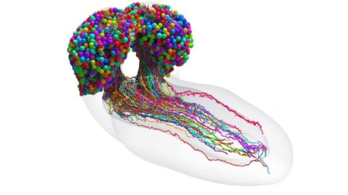
This team’s connectome of a fruit fly hatchling, the larva of Drosophila melanogaster , is the most complete and extensive map of a whole insect brain ever completed. It includes 3,016 neurons and each connection between them: 548,000.

Vogelstein said:
“It’s been 50 years and this is the first brain connectome. It’s a flag in the sand that we can do this.”
“Everything has been working until this.”
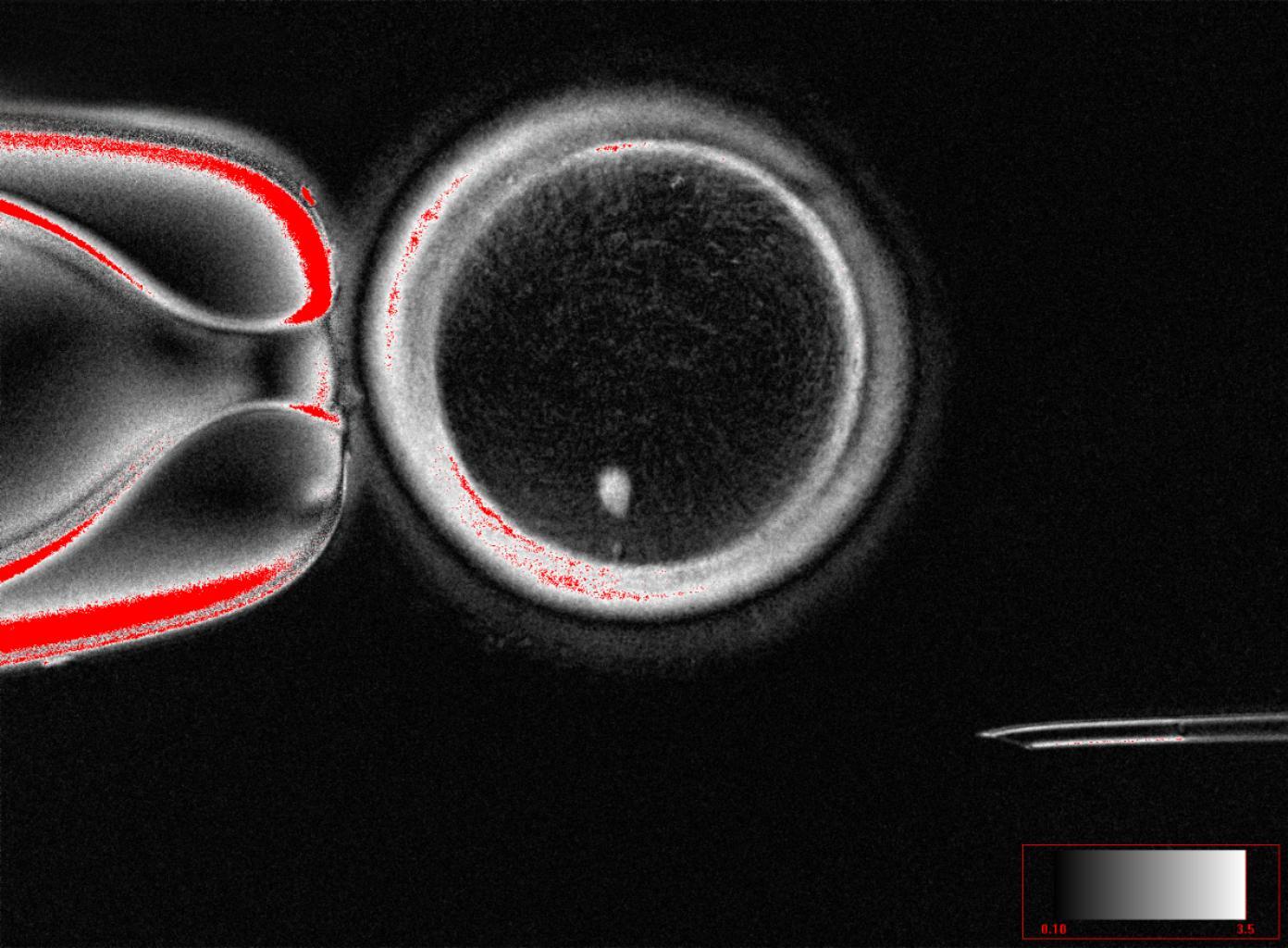
Mapping whole brains is difficult and time-consuming, even with the best modern technology. Obtaining a complete cellular-level image of a brain requires slicing the brain into hundreds or thousands of individual tissue samples, all of which have to be captured with electron microscopes before the painstaking process of reconstructing all those pieces, neuron by neuron, into a complete, accurate portrait image of a brain.
It took more than a decade to do that with baby fruit flies. The brain of a mouse is estimated to be one million times larger than that of a baby fruit fly, which means that the possibility of mapping anything resembling a human brain is not likely in the near future, perhaps not even in our lifetimes.
The team deliberately chose fruit fly larvae because, for an insect, the species shares much of its fundamental biology with humans, including a comparable genetic base. It also has rich learning and decision-making behaviors, making it a useful model organism in neuroscience. And for practical purposes, their brains can be imaged relatively compactly and their circuits can be reconstructed in a reasonable time frame.
Even so, the work took the University of Cambridge and Johns Hopkins 12 years. The imaging alone took about one day per neuron.
The Cambridge researchers created the high-resolution images of the brain and studied them manually to find individual neurons, rigorously tracing each one and linking their synaptic connections.
Cambridge delivered the data to Johns Hopkins, where the team spent more than three years using the original code they created to analyze brain connectivity. The Johns Hopkins team developed techniques to find clusters of neurons based on shared connectivity patterns and then analyzed how the information might propagate through the brain.
In the end, the entire team recorded every neuron and every connection, and classified each neuron according to the role it plays in the brain. They found that the most active circuits in the brain were those going to and from neurons in the learning center.
The methods developed by Johns Hopkins are applicable to any brain wiring project, and their code is available to anyone trying to map an even larger animal brain, Vogelstein said, adding that, despite the challenges, scientists are expected to tackle the mouse, possibly within the next decade. Other teams are already working on a map of the adult fruit fly brain.
Co-author Benjamin Pedigo, a PhD candidate in biomedical engineering at Johns Hopkins, hopes the team’s code can help reveal important comparisons between connections in the adult and larval brain. As connectomes are generated for more larvae and other related species, Pedigo hopes that his analysis techniques can lead to a better understanding of variations in brain wiring.
The fruit fly larvae work showed circuit characteristics that were strikingly reminiscent of prominent and powerful machine learning architectures. The team expects the ongoing study to reveal even more computational principles and potentially inspire new artificial intelligence systems.
Vogelstein said:
“What we learned about the code of fruit flies will have implications for the code of humans.”
“That’s what we want to understand: how to write a program that leads to a human brain network.”
COMPARTE ESTE ARTICULO EN TUS REDES FAVORITAS:

Esta entrada también está disponible en:
![]() Español
Español
Discover more from Cerebro Digital
Subscribe to get the latest posts sent to your email.