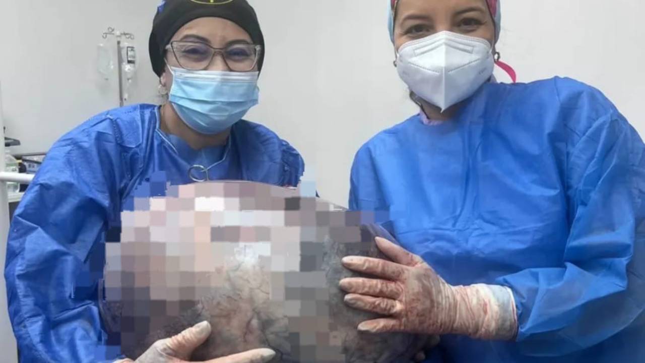

En un caso extremadamente raro, los médicos extirparon quirúrgicamente un feto del cerebro de una niña de 1 año. Los médicos lo encontraron después de que la niña mostrara un retraso en el desarrollo de las habilidades motoras, una circunferencia de la cabeza agrandada y una acumulación de líquido en el cerebro.
Feto extraído del cerebro de una niña

a “malformed diamniotic monochorionic twin,” meaning the fetuses had once shared the same placenta in the womb but had separate amniotic sacs—thin-walled, fluid-filled sacs surrounding developing fetuses. These types of twins come from the same fertilized egg, making them identical.
The anomaly where one fetus becomes enclosed by another is known as “fetus in fetu” or sometimes “parasitic twin.” The absorbed twin usually stops developing while the other continues growing.
The phenomenon occurs in about 1 in 500,000 live births; typically, the malformed fetus appears as a mass in the other’s abdomen, nestled behind tissues lining the abdominal wall. Here, however, the mass appeared in the host child’s head and likely arose very early in development, when the fertilized egg forms a cell cluster called a blastocyst.
It Had Developed Bones

“Se propone que el feto en feto intracraneal surja de blastocistos no separados”, lo que significa que los grupos de células que estaban destinados a crecer en dos individuos separados permanecieron pegados, escribieron los autores del informe del caso.
“The joined parts develop into the host fetus’s forebrain and envelop the other embryo during neural plate folding.” (The neural plate is a structure formed in early development that gives rise to the nervous system).
2020 case report in World Neurosurgery:
“Intracranial fetus in fetu is extremely rare with fewer than 20 reports published worldwide.”
The 1-year-old girl’s head brain scans revealed the fetus contained a spine and two leg bones (femur and tibia), and had spina bifida—a condition where part of the spinal cord remains exposed, rather than covered by back tissues, due to a developmental issue. Once removed, the fetal mass was also found to have “upper limb buds and digits.”
The brief report by Fudan University doctors, who performed the extraction, provides no details on the 1-year-old girl’s condition post-surgery.
COMPARTE ESTE ARTICULO EN TUS REDES FAVORITAS:

Esta entrada también está disponible en:
 Español
Discover more from Cerebro Digital
Subscribe to get the latest posts sent to your email.